Snapping Hip
“My hip pops and snaps and feels like it is popping out of place”
Last week I discussed the role of the hip labrum and the management of labral tears. We learned that labral tears can cause popping, clicking and snapping in the hip. Internal and external snapping hip, otherwise known as “Coxa Sultans Interna” and “Coxa Sultans Externa”, are other very common conditions of the hip that can cause popping sensations. This condition is seen in all types of athletes and in high intensity training exercises, but none as common as ballet dancers in which over 90% have this painless condition and is referred to as “Dancer’s Hip”. Have you ever performed repetitive leg raises or flutter kicks in the gym and with every repetition you can feel a reproducible pop or clunking sensation? Also, do you feel like you can pop your hip out of place by shifting your hip in a specific way that causes visible and audible clunking and popping? If you answered yes to either of these questions you may have internal or external snapping hip and in the next section I will discuss the pathophysiology that causes these phenomena and how to treat them.

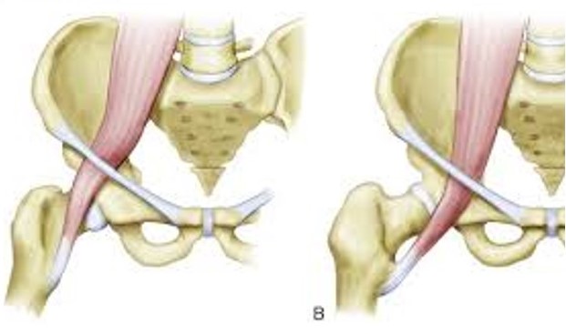
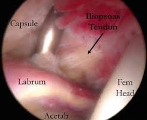
Internal snapping hip is a condition present in 10% of the normal population and as high as 25% in athletes and is caused by the hip flexor (iliopsoas tendon) snapping over the front rim of the hip socket (acetabulum). The vast majority of the time this is a painless snapping and treatment is not necessary. The causes of this problem are typically due to overuse, but it can also be due to anatomic variations of the hip that can cause irritation of the tendon. Most commonly the cause is a tight hip flexor but it also can be due to rubbing of the tendon over the bony rim of the pelvis (iliopectineal eminence) or over the front rim of the hip socket which are intimately located to the tendon. Different variations in shape of the socket can make internal snapping hip more common: femoroacetabular impingement (FAI-refer to this section for more information), retroversion of the socket (angled slightly to the back as opposed to the normal anteverted position), and subtle hip instability. Additionally, patients may experience painful snapping of the hip following a total hip replacement due to irritation of the tendon by the metal cup or liner of the socket. If the snapping becomes painful it is often because the tendon is inflamed (tendonitis) or the bursa sac deep to the tendon is enlarged (bursitis). Treatment for painful snapping is a systematic multimodal approach with avoidance of the aggravating activity and formal course of physical therapy for iliopsoas stretching, core, hip gluteal and groin muscle strengthening, and possibly dry needling. Additionally, anti-inflammatory medication (ibuprofen, naproxen) with rest is recommended. However, if pain persists despite these treatments, an ultrasound examination can confirm the diagnosis by visualizing the snapping and swelling around the tendon and can aid in injecting corticosteroid, or even platelet rich plasma (see PRP section) around the tendon or into the bursa for potential long-term relief in over 90% of patients. Cases that are recalcitrant to this treatment may be due to the fact that there is a high incidence of concomitant pathology on the inside of the ball and socket joint. Commonly associated intra-articular conditions that may need to be addresses at the time of surgery include labral tears (see the labral tear section for information), cartilage abnormality, or both which can be seen in hip impingement (FAI). When a patient comes to my office for evaluation I will perform a thorough physical exam looking for evidence of hip snapping and then assess for hip flexor tightness, core weakness, or muscle imbalances. I will also obtain a series of x-rays looking for structural abnormalities that may have led to the snapping. Dynamic ultrasound can be used in the office if needed to confirm the snapping while the patient is actively moving the hip and reproducing the pain. An MRI can be useful to evaluate the soft tissue structures of the hip and assess the labrum and cartilage. If conservative treatment does not improve the pain and an injection provided temporary relief, arthroscopic release/lengthening of the hip flexor can be performed where studies have shown no recurrent snapping and recreational and competitive athletes have returned to activity within 4 months from surgery with normal strength and function. The surgery is same day and takes 20-30 minutes depending on other conditions that may need to be addressed at the time of surgery. I utilize 2 small incisions (2-3mm) and complete the surgery using small camera and instruments. Recovery is a four-phase process and initially the patient will require protected weight bearing with crutches for 1-2 weeks and a hip brace can be utilized. Stationary bike is started immediately and patient can progress with return to jogging at 3 months. By 4.5 months most patients have restored strength and function.

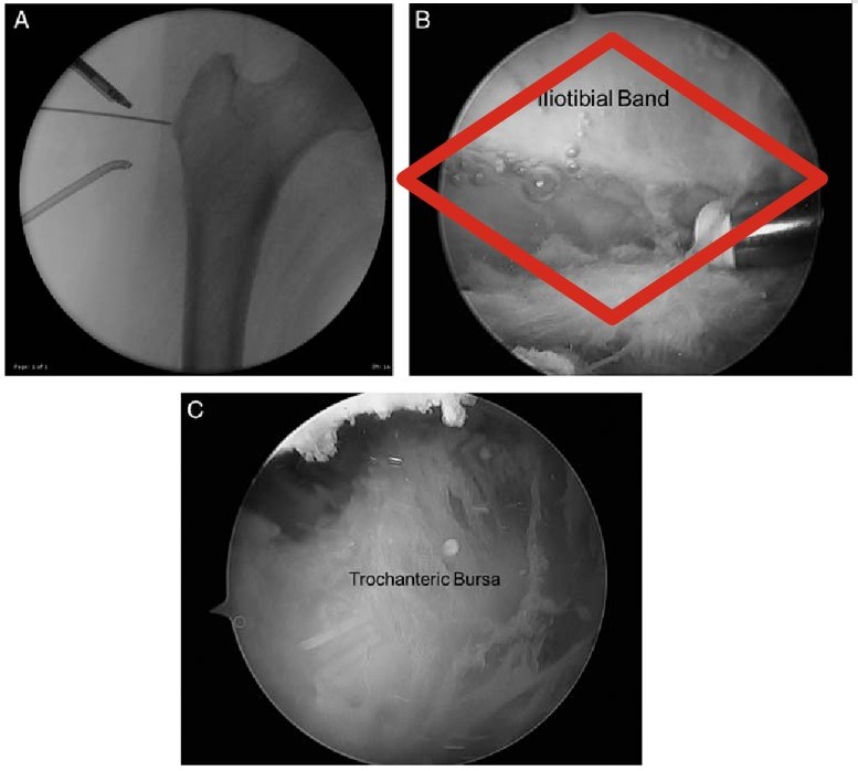
External Snapping hip is a condition caused by snapping of the iliotibial band (ITB) over the bony prominence on the outside of the hip known as the greater trochanter when the hip goes from a flexed to an extended position. The snapping in this condition is typically volitional and can be an extremely loud snap that can be seen and heard from across a room. Again, this condition is quite common and rarely does it cause pain. Patients can mistake this maneuver as their hip coming out of socket or shifting out of place. It is very uncommon for the hip to “come out of socket” as the hip joint is a highly constrained joint due to its bony shape and strong hip joint capsule. Physical therapy is the mainstay of treatment for this condition which focuses on ITB and gluteal maximus stretching, gluteal and core strengthening, and dry needling. X-rays will be obtained in clinic and a dynamic ultrasound can also be used to make the diagnosis. This condition can become painful due to bursitis in the area and can also lead to micro tearing of the gluteal muscles (hip abductors) that insert in that area. Weakness of the stabilizers of the hip may also be appreciated on physical exam. Corticosteroid and sometimes PRP can help alleviate the symptoms. If conservative management fails an MRI can help assess the bursitis in the area as well as look at the gluteal tendons to evaluate for tears that may require arthroscopic repair. If tears are present this may present as weakness of the stabilizers of the hip on physical exam. If surgery is indicated after the appropriate trial of conservative treatment fails, the management is to arthroscopically resect the portion of the ITB that snaps over the bone prominence. The structure is not detached as only a small window is created in it in order for the snapping to be alleviated. Rarely does the hip gluteal tendons need to be repaired at the same time. The surgery takes approximately 20-30 minutes and is performed through 2-3 minimally invasive arthroscopic incisions and utilizes a camera and small instruments. The rehabilitation follows the same 4 phase recovery as mentioned previously with a brief period of bracing and crutch use and typically patients return to full activity by 3-4 months. Studies have shown 98-100% return to pre-injury activity.

If you are experiencing symptoms consisted with snapping hip and would like to be seen please go to the “make an appointment” section of this site and request an appointment to be evaluated. I am confident that with the timely and correct diagnosis and the appropriate conservative treatment this condition can be improved without surgery. For the rare cases that require surgical attention the patient reported outcomes and return to sport activity are excellent.